Sketch And Label Of A Cross Section Of A Long Bone - Bone Human Anatomy - Draw and label the following structures as they appear using the 10x objective o bone marrow o bony trabeculae activity 5.2.3:
Sketch And Label Of A Cross Section Of A Long Bone - Bone Human Anatomy - Draw and label the following structures as they appear using the 10x objective o bone marrow o bony trabeculae activity 5.2.3:. Draw and label the following structures as they appear using the 10x objective o bone marrow o bony trabeculae activity 5.2.3: A long bone has a shaft and 2 ends. External circumferential lamellae, osteon, central canal, perforating canals, lacuna, canaliculi, concentric lamellae. Make sure learners follow all the criteria for a biological drawing. Sketch and label of a cross section of a long bone.
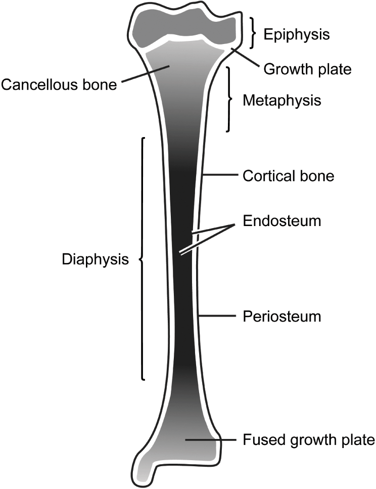
External circumferential lamellae, osteon, central canal, perforating canals, lacuna, canaliculi, concentric lamellae. In the space provided, draw a longitudinal section of a long bone and label the following parts: The diaphysis and the epiphysis. The diaphysis of a long bone is composed of bone tissue while the epiphysis is made of bone tissue. The diaphysis is the tubular shaft that runs between the proximal and distal ends of the bone.
Applied Basic Sciences Section 5 Postgraduate Orthopaedics from static.cambridge.org The little black spots are osteocytes. It suggests that the bone will have equal strength in all directions. Bone test anatomy and physiology 12 photos of the bone test anatomy and physiology anatomy and physiology bone lab test, anatomy and physiology bone markings test, anatomy and physiology bone practical test, anatomy and physiology bone tissue test, anatomy and physiology test on bone tissue, bone, anatomy and. The structure of a long bone allows for the best visualization of all of the parts of a bone (figure 1). The structure of a long bone allows for the best visualization of all of the parts of a bone ( figure 6.7 ). This is the long central shaft. Compact bone is very different from the other tissues you have seen. Terms in this set (3) epiphysis.
This slide contained a cross section of a very small bone, and you are looking at the entire thickness of the shaft of the bone.
Learners should accurately draw a long bone, resembling that in figure 6.24. This is the long central shaft. A long bone has a shaft and 2 ends. Smartdraw includes 1000s of professional healthcare and anatomy chart templates that you can modify and make your own. The diaphysis and the epiphysis. Osteons are oriented parallel to the diaphysis of the long bone. Related posts of cross section of a long bone bone test anatomy and physiology. Explain the functions of each of the labeled structures. The little black spots are osteocytes. The head of each end of a long bone consists largely of spongy bone and is covered with hyaline cartilage. Once we stop growing (between 18. While it is not as hard as compact bone, spongy bone plays an important role of protecting the marrow where blood cells are produced. Marks should be deducted for shading or colouring.
Proximal epiphysis, distal epiphysis, diaphysis, metaphysis, medullary cavity, epiphyseal line 2. The diaphysis is the tubular shaft that runs between the proximal and distal ends of the bone. A typical long bone shows the gross anatomical characteristics of bone. Make a pencil sketch and use markers or colored pencils to add details. Sketch and label of a cross section of a long bone.
1 19 Describe The Structure Of Bone And Label A Diagram Of A Typical Long Bone In Longitudinal Section Diagram Quizlet from o.quizlet.com Cross section of a long bone. A long bone has a shaft and 2 ends. The end of a growing tibia, cut lengthwise*. Marks should be deducted for shading or colouring. Smartdraw includes 1000s of professional. Plates of cartilage, also known as growth plates which allow the long bones to grow during childhood. Draw and label the following structures as they appear using the 10x objective o bone marrow o bony trabeculae activity 5.2.3: A typical long bone shows the gross anatomical characteristics of bone.
Sketch and label of a cross section of a long bone.
Explain the functions of each of the labeled structures. Growth in length of a bone occurs at the 4. Lamellar bone makes up the compact or cortical bone in the skeleton, such as the long bones of the legs and arms. Long bones have a thick outside layer of compact bone and an inner medullary cavity containing bone marrow. Anatomy of a long bone 1. 100x first focus in the compact decalcified bone (cb) on the left part of the image, you can see small dots, which are. This is an online quiz called label the long bone. Related posts of cross section of human bone diagram foot bone anatomy x ray. The ends of a long bone contain spongy bone and an epiphyseal line. While it is not as hard as compact bone, spongy bone plays an important role of protecting the marrow where blood cells are produced. Sketch and label a cross section of a bone. It suggests that the bone will have equal strength in all directions. Following is an idealized roadway typical section with the various elements identified.
You need to get 100% to score the 10 points available. Growth in length of a bone occurs at the 4. Slides have to be made this way because the matrix of bone is too hard to be cut with a knife as the other tissues are. Create a drawing of the bone section in your laboratory journal and label the areas listed above. 100x first focus in the compact decalcified bone (cb) on the left part of the image, you can see small dots, which are.
Chapter 6 Intro Skeletal System Diagram Quizlet from o.quizlet.com The structure of a long bone allows for the best visualization of all of the parts of a bone ( figure 6.7 ). In these labeled examples, a human femur is represented without identifying many of the unique characteristics that help differentiate the femur bone from other bones in the human body. Bone test anatomy and physiology 12 photos of the bone test anatomy and physiology anatomy and physiology bone lab test, anatomy and physiology bone markings test, anatomy and physiology bone practical test, anatomy and physiology bone tissue test, anatomy and physiology test on bone tissue, bone, anatomy and. Related posts of cross section of human bone diagram foot bone anatomy x ray. End of a long bone. Following is an idealized roadway typical section with the various elements identified. Explain the functions of each of the labeled structures. Draw and label a longitudinal section of a long bone.
A long bone has two parts:
Cross section of a long bone. Label the haversian canal, osteocyte (mature bone cell) in lacuna, and canaliculi. A typical long bone shows the gross anatomical characteristics of bone. There is a printable worksheet available for download here so you can take the quiz with pen and paper. You need to get 100% to score the 10 points available. Growth in length of a bone occurs at the 4. Compact bone is very different from the other tissues you have seen. A long bone has two parts: Make sure learners follow all the criteria for a biological drawing. In these labeled examples, a human femur is represented without identifying many of the unique characteristics that help differentiate the femur bone from other bones in the human body. Slides have to be made this way because the matrix of bone is too hard to be cut with a knife as the other tissues are. Anatomy of a long bone 1. The diaphysis is the tubular shaft that runs between the proximal and distal ends of the bone.


0 Komentar